Physiol. Meas. 27 (2006) 817–827 doi:10.1088/0967-3334/27/9/005
Impedance cardiography revisited
G Cotter1, A Schachner2, L Sasson2, H Dekel2 and Y Moshkovitz3
1 Divisions of Clinical Pharmacology and Cardiology, Duke University Medical Center, Durham,
NC, USA
2 Angela & Sami Sharnoon Cardiothoracic Surgery Department, Wolfson Medical Center, Israel
3 Department of Cardiac Surgery, Assuta Hospital, Petah Tikva, Israel
E-mail: gad.cotter@duke.edu
Received 7 February 2006, accepted for publication 9 June 2006
Published 5 July 2006
Online at stacks.iop.org/PM/27/817
Abstract
Previously reported comparisons between cardiac output (CO) results in
patients with cardiac conditions measured by thoracic impedance cardiography
(TIC) versus thermodilution (TD) reveal upper and lower limits of agreement
with two standard deviations (2SD) of approximately ±2.2 l min−1, a 44%
disparity between the two technologies. We show here that if the electrodes
are placed on one wrist and on a contralateral ankle instead of on the chest, a
configuration designated as regional impedance cardiography (RIC), the 2SD
limit of agreement between RIC and TD is ±1.0 l min−1, approximately
20% disparity between the two methods. To compare the performances of
the TIC and RIC algorithms, the raw data of peripheral impedance changes
yielded by RIC in 43 cardiac patients were used here for software processing
and calculating the CO with the TIC algorithm. The 2SD between the TIC
and TD was ±1.7 l min−1, and after annexing the correcting factors of the
RIC formula to the TIC formula, the disparity between TIC and TD further
declined to ±1.25 l min−1. Conclusions: (1) in cardiac conditions, the RIC
technology is twice as accurate as TIC; (2) the advantage of RIC is the use
of peripheral rather than thoracic impedance signals, supported by correcting
factors.
Keywords: cardiac output measurements, thoracic bioimpedance, whole-body
bioimpedance, impedance cardiography
Introduction
Three basic technologies are currently in use for impedance cardiography (ICG): (1) the
thoracic ICG (TIC), where the electrodes are placed on the root of the neck and the lower
part of the chest, being the dominant method in the market (Patterson et al 1964, Kubicek
0967-3334/06/090817+11$30.00 © 2006 IOP Publishing Ltd Printed in the UK 817
818 G Cotter et al
et al 1966, 1974); (2) the whole-body ICG (ICGWB), where four pairs of electrodes are used,
one pair on each limb (Tischenko 1973, Koobi et al 1999); (3) the regional ICG (RIC), a
technology which is used by the NICaS (noninvasive cardiac system). In this technology,
which is the subject of this report, only two pairs of electrodes are used, performing best
when placed on one wrist and on the contralateral ankle (Cohen et al 1998, Cotter et al 2004,
Torre-Amione et al 2004).
Two comprehensive reviews of the literature on clinical experience in measuring the
cardiac output (CO) by TIC determined that in patients with cardiac conditions the TIC-CO
results are unreliable (Raaijmakers et al 1999, Handelsman 1991). According to Patterson
(1985) andWang et al (2001), a number of sources in the chest, such as the lungs, vena cava, and
systemic and pulmonary arterial vasculatures, generate systolic impedance reductions, while
the heart generates signals of increased impedance. In addition to thesemultifarious sources of
Z,4 variations in the electrical conductivities between the sources of impedance changes and
the TIC electrodes (Kim et al 1988, Kauppinen et al 1998), and between the various impedance
sources (Wtorek 2000) have been described. These model experimentations indicated that
the thoracic
Z is not a reliable signal for calculation of the SV due to the multiple sources
of dZ/dt (Kim et al 1988, Wang and Patterson 1995, Kauppinen et al 1998, Wtorek 2000),
providing the explanations for the above-mentioned unsatisfactory clinical results obtained by
TIC (Raaijmakers et al 1999, Handelsman 1991).
In this report, an attempt is made to define the differences between the peripheral and
thoracic impedance signals, and based on this, to explain the differences in the performance
of RIC and TIC.
As we are capable of saving raw data from the wrist–ankle (peripheral) impedance
signals, we were able to use the peripheral impedance waveforms and base impedance values
to calculate stroke volumes, using various algorithms that have been associated with TIC
calculations. This enabled us to prove that (1) the performance of RIC is twice as accurate
as reported TIC results; (2) the reasons for this are as follows: (a) the impedance changes
which are yielded by the limb electrodes are more suitable than the impedance changes of
the thoracic electrodes for calculating the stroke volume and (b) the use of properly designed
coefficients improved the accuracy of the CO results by at least an additional 25%.
Methods
The data for this project were gathered from two patient series. In both, comparisons were made
between cardiac output results measured by the NICaS versus thermodilution. One series,
which was studied in hospital A, consisted of 30 patients who were studied immediately upon
arrival at the ICU following an open heart operation. In 11 (36%), despite the intravenous
administration of adrenalin, cardiac index (CI) was lower than 2.5 l min−1 m−2. The second
series included 13 cases of acute heart failure that were studied in hospital B. CI was lower
than 2.5 l min−1 m−2 in seven (54%), and in the combined group of 43 cases of the two
hospitals, it was lower than 2.5 l min−1 m−2 in 18 (43%).
The purpose of this study was to use peripheral impedance waveforms to calculate stroke
volume by means of four different ICG algorithms and to compare each of these SV values
with the thermodilution SV result.
Of the 55 and 31 studies conducted in hospitals A and B, respectively, raw data were
successfully retrieved from only the last 30 consecutive patients of hospital A and the last 13
4 In the ICGWB and RIC, where the impedance changes are depicted in the periphery, the impedance value is
automatically converted into the real parts (R0 and
R) of the measured impedance signals (Lamberts et al 1984).
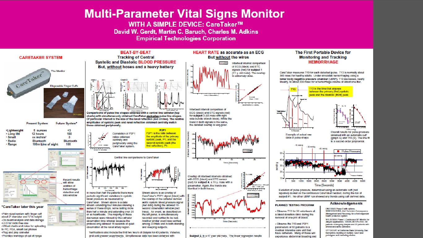
Markers of temporal changes in central blood volume are required to non-invasively detect hemorrhage and the onset of hemorrhagic shock. Recent work suggests that pulse pressure may be such a marker. A new approach to tracking blood pressure, and pulse pressure specifically is presented that is based on a new form of pulse pressure wave analysis called Pulse Decomposition Analysis (PDA). The premise of the PDA model is that the peripheral arterial pressure pulse is a superposition of five individual component pressure pulses, the first of which is due to the left ventricular ejection from the heart while the remaining component pressure pulses are reflections and re-reflections that originate from only two reflection sites within the central arteries. The hypothesis examined here is that the PDA parameter T13, the timing delay between the first and third component pulses, correlates with pulse pressure.
T13 was monitored along with blood pressure, as determined by an automatic cuff and another continuous blood pressure monitor, during the course of lower body negative pressure (LBNP) sessions involving four stages, -15 mmHg, -30 mmHg, -45 mmHg, and -60 mmHg, in fifteen subjects (average age: 24.4 years, SD: 3.0 years; average height: 168.6 cm, SD: 8.0 cm; average weight: 64.0 kg, SD: 9.1 kg).
Results
Statistically significant correlations between T13 and pulse pressure as well as the ability of T13 to resolve the effects of different LBNP stages were established. Experimental T13 values were compared with predictions of the PDA model. These interventions resulted in pulse pressure changes of up to 7.8 mmHg (SE = 3.49 mmHg) as determined by the automatic cuff. Corresponding changes in T13 were a shortening by -72 milliseconds (SE = 4.17 milliseconds). In contrast to the other two methodologies, T13 was able to resolve the effects of the two least negative pressure stages with significance set at p < 0.01.
Conclusions
The agreement of observations and measurements provides a preliminary validation of the PDA model regarding the origin of the arterial pressure pulse reflections. The proposed physical picture of the PDA model is attractive because it identifies the contributions of distinct reflecting arterial tree components to the peripheral pressure pulse envelope. Since the importance of arterial pressure reflections to cardiovascular health is well known, the PDA pulse analysis could provide, beyond the tracking of blood pressure, an assessment tool of those reflections as well as the health of the sites that give rise to them.
Pulse Decomposition Algorithm and Intra-Arterial
Catheters in ICU Patients
Introduction
The object of the work presented here was to validate a new approach to tracking blood pressure that is
based on the pulse analysis of the peripheral arterial pressure pulse. The approach, referred to as the
Pulse Decomposition Analysis (PDA) model, goes beyond traditional pulse analysis by invoking a
physical model that comprehensively links the components of the peripheral pressure pulse
envelope with two reflection sites in the centralarteries. The first reflection site is the juncture
between thoracic and abdominal aorta, which is marked by a significant decrease in diameter and a
change in elasticity. The second site arises from the juncture between abdominal aorta and the common
iliac arteries.
The PDA model integrates and goes beyond the findings of a number of studies that have confirmed
the existence of the two reflection sites. [Kriz 2008,
Latham 1985] A consequence of these reflection sites are two reflected arterial pressure pulses, referred
to as component pulses, which counter-propagate to the direction of the single arterial pressure pulse,
due to left ventricular contraction, that gave rise to them. The scenario, sketched in Figure 1, has been
described in detail elsewhere. [Baruch 2011] In the arterial periphery, and specifically at the radial or
digital arteries, these reflected pulses, the renal reflection pulse (P2, also known as the second systolic
pulse) and the iliac reflection pulse (P3), arrive with distinct time delays. In the case of P2 the delay is
typically between 70 and 140 milliseconds, in the case of P3 between 180 to 450 milliseconds.
Quantification of physiological parameters is accomplished by extracting pertinent component pulse
parameters. In the case of the beat-by-beat tracking of blood pressure the PDA model’s predictions and
previous experimental studies have shown that two pulse parameters are of particular importance. The
ratio of the amplitude of the renal reflection pulse (P2) to that of the primary systolic pulse (P1) tracks
changes in beat-by-beat systolic pressure. The time difference between the arrival of the primary
systolic (P1) pulse and the iliac reflection (P3) pulse, referred to as T13, tracks changes in arterial pulse
pressure.
Patients and Methods
In these experiments, approved by the University of Virginia Medical Center Review Board, the arterial
blood pressures of patients (23 m/11 f, mean age: 44.05 y, SD: 13.9 y, mean height: 173.3 cm, SD: 9.4
cm, mean weight: 95.3 kg, SD: 27.4 kg) hospitalized in the University of Virginia Medical Intensive Care
Unit (MICU) were monitored using radial intra-arterial catheters, while the CareTaker system collected
pulse line shapes at the lower phalange of the thumb. The overlap recording sessions with both
monitoring systems were scheduled for four hours but were frequently shorter because of medical
procedures. BedMaster (Excel Medical, Jupiter, FL) hardware and software was used to digitize and
record intra-arterial waveform data from the GE Unity network at a sample rate of 240 Hz.
Consent for data collection was obtained for 60 patients, and 34 successful data overlaps between
arterial catheter and CareTaker were obtained. Eleven arterial data sets were lost because of a
programming error in the BedMaster software, causing data to be overwritten when the overall arterial
catheter recording session extended beyond one day after the CareTaker/arterial catheter overlap
recording window. In 4 cases the BedMaster system was found to have been inoperative during the
recording window. In 4 cases the arterial catheter recordings did not include the overlap recording
window for unknown reasons, while in 4 additional cases the arterial catheter failed. Two cases involved
such substantial movement artifacts due to movement, extubation etc. that neither system was able to
obtain valid recordings. In one case the CareTaker device became accidently disconnected early in the
session.
A. CareTaker Device and PDA model
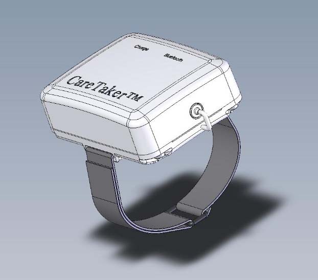
The hardware platform, which is the Care-Taker device (Empirical Technologies Corporation,
Charlottesville, Virginia) the model, and the algorithm implementation have been described in detail
elsewhere [Baruch].
B. Statistical analysis
We present regression coefficients and linear fits between arterial catheter blood pressures and the PDA
parameters P2P1 and T13 for an individual patient, histogram distributions of the slopes of the linear fits
for individual patients, as well as overall linear fit-based comparisons between central catheter blood
pressures and blood pressures obtained from the PDA parameters using a single set of conversion
constants. Bland-Altman comparisons of the two sets of blood pressures are also provided.
C. Data Inclusion
Data were excluded from analysis on the following basis:
In the case of the arterial catheter data the criteria for exclusion were as follows: A. visual inspection of
the data was used to identify sections of obvious catheter failure, characterized by either continuous or
pervasive intermittent analog/digital readings of -/+32768. B. sections contaminated by excessive
motion artifact were identified as such if the peak detection algorithm was no longer able to identify
FactorTime (Seconds)
heart beats, as evidenced by inspection of the resulting implausible and discontinuous inter-beat
interval spectrum.
In the case of the CareTaker data a Fourier spectral analysis approach was used to establish a
signal/noise factor (SNF) to identify poor quality data sections. To this end the standard deviations and
integrated amplitudes of different spectral bands were calculated. The band associated with the
physiological signal was chosen from 1-10 Hz, based on data by the authors and published results by
others. The signal strength associated with this band was compared with those of the 100-250 Hz
frequency band, which is subject to ambient noise but contains no physiologically relevant signal. The
spectrogram (top graph) displayed in figure 10 provides a motivation. Data sections characterized by low
noise feature low high-frequency spectral amplitudes and high low-frequency spectral amplitudes as
well as “structured” physiological bands, i.e. significant amplitude variations between the harmonic
bands. This motivates the analytical form of the SNF parameter, which is established by taking the ratio
A-line DiastoleTime (Seconds)
of the standard deviation of the 1-10 Hz band to the product of the integrated band strengths of the 1-
10 Hz and 100-250 frequency bands. The bottom graph of Figure 2 displays the SNF parameter for the
raw CareTaker data presented in the center graph of Figure 2. Data sections with an SNF below 80 were
excluded from the analysis.
Results
The overlap of the CareTaker data streams and the central catheter data streams was established, after
an initial alignment based on data collection systems clocks, by matching time-based inter-beat interval
spectra obtained via pulse detection obtained from both data streams. Figure 3 presents an example of
such an overlap for patient 21. The figure presents both an overall overlay of the data streams, as well
as a 70 second expanded section.
The linear model used to perform the conversion of the P2P1 parameters to systolic blood pressure for
patient 21 was (140 × P2P1 (unitless ratio) + 59.2), while the corresponding linear conversion for the T13
parameter to pulse pressure was (0.1 × T13 (milliseconds) + 13.6). Both the offsets and the slope factors
were patient-specific and obtained by chi^2 minimization of the fit of both data streams. The details of
obtaining the slope factor and the offset for individual patients, as well as the physiological relevance of
the slope factor regarding arterial stiffness, are discussed later.
In figures 9 and 10 we present the correlation and Bland-Altman results for the interbeat interval data
for patient 21, which was presented in Figure 2. The data presented in the Bland-Altman plot of figure 9
demonstrates the resolution limits of the data streams, which presents itself as diagonally-running
striations that are visible in the data. Had the data rate in both data streams been equal, the striations
would run at 45 degrees. In the case of the CareTaker data, the acquisition rate was 500 Hz, or a data
point spacing of 2.0 milliseconds, while the catheter-based data was collected at 240 Hz, corresponding
to a data point spacing of 4.16 milliseconds. Consequently the slope of the striations is 0.48, the ratio of
lower to higher data acquisition rate.
Overall Blood Pressure Results
The overall results of the study are presented as blood pressure correlations and Bland-Altman graphs in
figures 11 through 14. The data comparisons are based on two-second averages of 40-minute sections
from each patient. This approach is an established procedure that has been used in previous
presentations of comparative blood pressure data by others. 1 One important benefit of this approach is
that it gives equal weight to the data set of each patient
1 Martina JR et. al., Noninvasive continuous arterial blood pressure monitoring with Nexfin, Anesthesiology. 2012
May;116(5):1092-103.
Average Inter-beat Interval (Seconds))
mean: -0.056 millisecondsSD: 6.0 milliseconds
larger standard deviation observed here is affected by two factors: 1. the comparatively low sampling
rate of the catheter signal, 240 Hz corresponding to 4.2 milliseconds, and 2. the significantly broader
structure of the blood pressure pulse compared to the EKG signal. The first limits the temporal
resolution of peak detection, while the second limits the threshold accuracy with which a given peak can
be detected relative to other peaks, likewise introducing uncertainty into the temporal accuracy.


