顕微鏡の使い方はこちら(How to use microscope)↓
中学全学年理科① 顕微鏡の使い方 JHS biology ~Microscope~
【目的】
身の回りの生物の表面のつくりを観察して、どのような特徴があるのか調べる。
【Goal】
Observe the surface structure of organisms around us and study what characteristics they have.
【準備物】
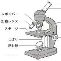
光学顕微鏡
スライドガラス
カバーガラス
ピンセット
柄つき針
ろ紙
カッターナイフ(またはカミソリ)
スポイト
タマネギ・トマト・レタスなど(タマネギがおすすめ)
【Preparation】
Microscope
microscope slide
cover glass
tweezers
needle with a handle
filter paper
box cutter kife
pipette
onion, tomato, lettuce etc. (recommend : onion)
【方法】
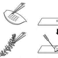
① タマネギの薄皮にカッターナイフで5mm四方の切り込みを入れ、ピンセットで薄皮をはぎ取る。
② 切り取った薄皮をスライドガラスの上に置き、スポイトで水を一滴垂らす。
③ 柄つき針で支えながら、ピンセットで薄皮の上にカバーガラスを静かに下ろし、プレパラートを作る。
④ 100倍程度の低倍率で観察したあと、400〜600倍の高倍率で観察する。
【Process】
① Cut the skin of onion (inside) as 5mm square and pick the skin up with tweezers.
② Put the piece of skin on microscope slide and put a drop of water with a pipette on it.
③ While supporting the cover glass with a needle with a handle, match the edge of it and the edge of the water.
Lower the cover glass on the skin of Onion with tweezers to prevent air from entering (preparation).
④ Observe with lower magnification (×100), later higher magnification (×400~600).


【結果】
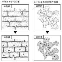
どの生物のからだも、小さな部屋のようなものが多数見られる。
【Results】
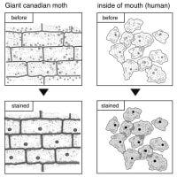
We can observe like some small "room"s in the body of any organisms.
【考察】
生物の体の共通点として「小さな部屋のようなもの」が集まってできている点が挙げられる。
【Study】
Common structure : the body of organisms is consist of many small "room"s.





※コメント投稿者のブログIDはブログ作成者のみに通知されます