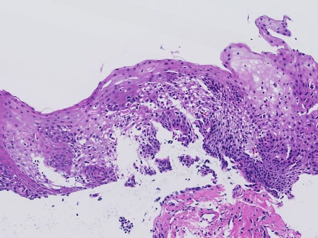
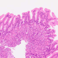
何度もみたことあるような組織像ですが(写真クリック!)、Lymphocytic esophagitis(LyE)という診断は日本ではあまり馴染まれていません。
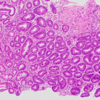
外国人病理医仲間達の会話を聞いていて、この病名がよく呟かれていたので、気になっていました。内視鏡的・臨床的にEosinophilic esophagitis(EoE)を疑って生検された検体です(胸部食道)。好酸球浸潤はほとんどなく、リンパ球ばっかりの浸潤です。しかし、見出し写真と下の写真の一枚目をみると、Civatte body??があるようにみえ、lichenoid esophagitisと言われるかもしれません。
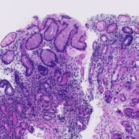
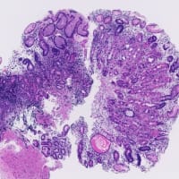
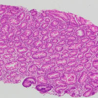
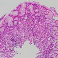
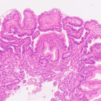
その他の生検像(いずれも胸部食道)です(下記、小写真クリックください)。
はたしてdistinctな病名になるかどうか??参考文献は(PMID: 30370453)
This is an image of an esophageal biopsy as often seen in routine gastrointestinal pathology work (click on the picture!). The term of the disease "Lymphocytic esophagitis" is not very familiar in Japan.
This is the name of the disease I am concerned about, since it has been often talked about by my close foreign GI-pathologists. This specimen was biopsied with endoscopic and clinical suspicion of eosinophilic esophagitis (EoE) (taken from the thoracic esophagus). There is almost no eosinophilic infiltrate, and the infiltrate is almost all lymphocytes. If you look closely, however, it could be regarded as "lichenoid esophagitis" because of some “civatte bodies?” seen in the headline photo and in the first photo below.
Other biopsy images (also taken from the thoracic esophagus) (click on the small pictures below).
Will this be a distinct disease? For references (PMID: 30370453)


隣国の病理学会に行って来ました。大阪から2時間弱なので1泊2日です。

胃炎の成り立ち-内視鏡診断のこれまで、これから
外国人病理医仲間達の会話を聞いていて、この病名がよく呟かれていたので、気になっていました。内視鏡的・臨床的にEosinophilic esophagitis(EoE)を疑って生検された検体です(胸部食道)。好酸球浸潤はほとんどなく、リンパ球ばっかりの浸潤です。しかし、見出し写真と下の写真の一枚目をみると、Civatte body??があるようにみえ、lichenoid esophagitisと言われるかもしれません。
その他の生検像(いずれも胸部食道)です(下記、小写真クリックください)。
はたしてdistinctな病名になるかどうか??参考文献は(PMID: 30370453)
This is an image of an esophageal biopsy as often seen in routine gastrointestinal pathology work (click on the picture!). The term of the disease "Lymphocytic esophagitis" is not very familiar in Japan.
This is the name of the disease I am concerned about, since it has been often talked about by my close foreign GI-pathologists. This specimen was biopsied with endoscopic and clinical suspicion of eosinophilic esophagitis (EoE) (taken from the thoracic esophagus). There is almost no eosinophilic infiltrate, and the infiltrate is almost all lymphocytes. If you look closely, however, it could be regarded as "lichenoid esophagitis" because of some “civatte bodies?” seen in the headline photo and in the first photo below.
Other biopsy images (also taken from the thoracic esophagus) (click on the small pictures below).
Will this be a distinct disease? For references (PMID: 30370453)


隣国の病理学会に行って来ました。大阪から2時間弱なので1泊2日です。

胃炎の成り立ち-内視鏡診断のこれまで、これから






韓国の学会でサインをいただいた
Seegene医療財団のSanghwa Leeともうします.
Lymphocytic esophagitis...
何度もみたことがあると思いますが
単純に Chronic esophagitisと診断していました.
今日も新しいものを教えてもらいました.
ありがとうございます.
처음으로 제 책에 사인을 했어요.