
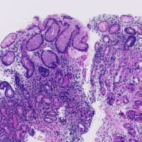
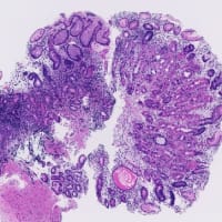
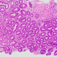
免疫チェックポイント阻害薬によるirAE( immune-related adverse events)腸炎と考えられた大腸生検組織です(写真クリック!)。
"Histolopathologic features of colitis due to immunotherapy with anti-PD-1 antibodies"というタイトルの論文(Greg先生チームによるAJSP, 2017)を読むと、irAE腸炎は
1) Active colitis with apoptosis
2) Lymphocytic colitis
に二大別されています。
1)はIBD(特にUC)に類似した組織像を示しますが、微妙に異なる点がいくつか指摘されています(ここでは省略)。
本例は2)相当です。陰窩の歪みや腺管密度低下はありませんが、粘膜固有層全層性に中等度のリンパ球・形質細胞浸潤がみられ好酸球・好中球が混じます。表層部上皮内リンパ球(IEL)が増多しています。うっすらとcollagen bandらしきものも形成されつつあります。

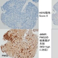
免疫染色を追加しますと(クリック!)、上皮内リンパ球はもっぱらCD8+Tリンパ球で、上皮細胞100個あたり20個は軽く越えています。
Microscopic colitisの組織像については、海外自主研修の学生がいつもお世話になっているグラーツ(墺太利)の先生方による"Histology of microscopic colitis"というタイトルのレビュー(Histopathology, 2015)がわかりやすいです。
This is a colon biopsy tissue considered to be IrAE (immune-related adverse events) colitis (click the photo!), due to an immune checkpoint inhibitor
According to "Histolopathologic features of colitis due to immunotherapy with anti-PD-1 antibodies" (AJSP, 2017 by Dr. Greg's team), irAE colitis is categorized into
1) Active colitis with apoptosis
2) Lymphocytic colitis
1) shows histopathological findings similar to those of IBD (especially UC), but some subtle differences have been pointed out.
This example is equivalent to 2). There is neither distortion of the crypts nor decrease in duct density, but moderate lympho-plasmocytic infiltration is observed in the lamina propria, and eosinophils/neutrophils are mixed. Intraepithelial lymphocytes (IELs) are increasing, positive for CD8. Mild thickening of collagen band is also seen.
Regarding the histopathological image of microscopic colitis, I recommend a review article (Histopathology, 2015) entitled "Histology of microscopic colitis" by a team of Graz , who kindly takes care of our medical students every summer.

大阪モノレール沢良宜駅
Osaka Monorail, Sawaragi Station
"Histolopathologic features of colitis due to immunotherapy with anti-PD-1 antibodies"というタイトルの論文(Greg先生チームによるAJSP, 2017)を読むと、irAE腸炎は
1) Active colitis with apoptosis
2) Lymphocytic colitis
に二大別されています。
1)はIBD(特にUC)に類似した組織像を示しますが、微妙に異なる点がいくつか指摘されています(ここでは省略)。
本例は2)相当です。陰窩の歪みや腺管密度低下はありませんが、粘膜固有層全層性に中等度のリンパ球・形質細胞浸潤がみられ好酸球・好中球が混じます。表層部上皮内リンパ球(IEL)が増多しています。うっすらとcollagen bandらしきものも形成されつつあります。

免疫染色を追加しますと(クリック!)、上皮内リンパ球はもっぱらCD8+Tリンパ球で、上皮細胞100個あたり20個は軽く越えています。
Microscopic colitisの組織像については、海外自主研修の学生がいつもお世話になっているグラーツ(墺太利)の先生方による"Histology of microscopic colitis"というタイトルのレビュー(Histopathology, 2015)がわかりやすいです。
This is a colon biopsy tissue considered to be IrAE (immune-related adverse events) colitis (click the photo!), due to an immune checkpoint inhibitor
According to "Histolopathologic features of colitis due to immunotherapy with anti-PD-1 antibodies" (AJSP, 2017 by Dr. Greg's team), irAE colitis is categorized into
1) Active colitis with apoptosis
2) Lymphocytic colitis
1) shows histopathological findings similar to those of IBD (especially UC), but some subtle differences have been pointed out.
This example is equivalent to 2). There is neither distortion of the crypts nor decrease in duct density, but moderate lympho-plasmocytic infiltration is observed in the lamina propria, and eosinophils/neutrophils are mixed. Intraepithelial lymphocytes (IELs) are increasing, positive for CD8. Mild thickening of collagen band is also seen.
Regarding the histopathological image of microscopic colitis, I recommend a review article (Histopathology, 2015) entitled "Histology of microscopic colitis" by a team of Graz , who kindly takes care of our medical students every summer.

大阪モノレール沢良宜駅
Osaka Monorail, Sawaragi Station






※コメント投稿者のブログIDはブログ作成者のみに通知されます